About this pathway
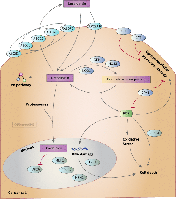
The anthracycline doxorubicin (DOX), a metabolite of Streptomyces peucetius var. caesius [Article:5365804], is a chemotherapeutic agent developed in the 1970s [Article:17652804] that is used in the treatment of a wide range of cancers, including non-Hodgkin's and Hodgkin's lymphoma, multiple myeloma, lung, ovarian, gastric, thyroid, breast, sarcoma, and pediatric cancers [Articles:17652804, 1462166]. This pathway shows the pharmacodynamics of doxorubicin in a stylized cancer cell and depicts candidate pharmacogenes.
Several mechanisms have been proposed to explain DOX antitumor activity [Article:10075079]. Here we describe 2 major mechanisms: the intercalation into DNA, leading to inhibition of the DNA synthesis or poisoning of topoisomerase II (TOP2A); and generation of free radicals, leading to DNA and cell membrane damage. We also show some pharmacokinetics (for more details see Doxorubicin PK Pathway) and depict transporters associated with drug resistance in cancer cell lines.
Intercalation into DNA
Anthracyclines are known to intercalate into DNA in vitro, and several crystal structures of complexes of DNA with DOX exist (see for example PDB entries 151D [Article:8142363] and 1P20 [Article:12717724]). In early in vitro studies, DOX was shown to cause DNA breaks [Article:566561] and to interfere with DNA synthesis [Articles:1277199, 1089410]. Other work has shown that the DNA-DOX interaction is related to the poisoning of topoisomerase II (TOP2A) [Articles:2551497, 6093249], but not topoisomerase I [Articles:2164630, 10385686]. Translocation of DOX into the nucleus is thought to occur via binding to proteasomes [Article:11289116]. Subsequent TOP2A poison-mediated cytotoxicity is postulated to involve the mismatch repair genes MSH2 and MLH1 [Article:11477562] because loss of DNA mismatch repair function results in resistance to doxorubicin [Articles:11477562, 9514047].
Topoisomerase II-mediated DNA damage is followed by cell death [Article:11172690]. TP53, a gene that is a major player of the DNA-damage response and apoptosis [Article:11790556], has been implicated in this DOX-apoptosis pathway. Several studies have shown an upregulation of TP53 occurs with anthracycline treatment [Articles:12739000, 10914720], and ERCC2 and TP53 have been shown to functionally interact in a p53-mediated apoptotic pathway with DOX treatment in lymphoblastoid cell lines [Article:10467415]. However, the actual involvement of p53 in DOX-induced apoptosis has been debated by other researchers [Article:11172690].
Generation of free radicals
DOX can undergo a one-electron reduction by several oxidoreductases to form a DOX-semiquinone radical [Article:2555273]. These enzymes include mitochondrial NADH dehydrogenases present in the sarcoplasmic reticulum and mitochondria: NDUFS2,3,7 EC 1.6.99.3 [Articles:12688675, 2850270, 9618942] as well as cytosolic enzymes NAD(P)H dehydrogenase (NQO1) [Article:12688675], xanthine oxidase (XDH EC 1.2.3.2) [Articles:12688675, 1911046] and endothelial nitric oxide synthase (NOS3) [Article:9333325]. Re-oxidation of the DOX-semiquinone radical back to DOX by leads to the formation of reactive oxygen species (ROS) and hydrogen peroxide [Article:9576481]. ROS, causing oxidative stress, can be deactivated by glutathione peroxidase, catalase and superoxide dismutase [Article:12751786].
Some researchers have related DOX free radical formation to cytotoxicity; these studies relate DOX cellular resistance to enzymes deactivating ROS. DOX has been shown to promote apoptosis in DOX-treated endothelial cells and myocytes through the formation of ROS and hydrogen peroxide [Article:12139490] via the activation of NF-kB (NFKB1, p50). But, activation of NF-kB blocked apoptotic cell death DOX-treated cancer cells [Article:12139490], indicating a possible different mechanism for cytotoxicity and cardiotoxicity. These researchers, and others, argue that the role of free radical formation is primarily related to cardiotoxicity and not cytotoxicity [Article:10075079], in part, because the use of an iron chelator, dexrazoxane, that binds the iron involved in the free radical formations, demonstrates cardioprotective properties without impacting clinical outcome [Articles:15038979, 9777314, 9193324, 9193323, 18425895].
Resistance
While DOX is a valuable clinical antineoplastic agent, in addition to problems with cardiotoxicity, resistance is also a problem limiting its utility [Articles:2982511, 1462166]. The mechanism of resistance is thought to involve, in particular, ABCB1 (MDR1, Pgp) and ABCC1 (MRP1) as well as other transporters.
In general, ABCB1 confers resistance by acting as an ATP-dependent drug efflux pump causing increased drug efflux [Article:8763334] via altered or increased expression [Articles:8763334, 9073310]. Cytotoxicity of DOX increases with inhibition of ABCB1 [Articles:1352877, 15788683]. Human ABCC1, originally cloned from a DOX-selected cancer cell line [Article:1360704] confers resistance to anthracyclines. Various studies on DOX-resistant cell lines have shown that resistance can be overcome via an inhibition of ABCB1, ABCC1 and ABCC2. [Articles:12370750, 15164094, 7214365, 1352877, 3180056, 17704753, 11172691, 7954421].
Studies have also shown an association between resistance and activity of other transporters. For example, RALBP1 activity was shown to be 2 times higher in a DOX-sensitive cell line versus DOX resistant cell line [Article:12527936]. In a study of a panel of lung cancer cell lines, a correlation between the DOX semiquinone levels and proteins levels for ABCC3 and ABCG2 was demonstrated [Article:11410522]. Glutathione transferase activity was found to be greater in DOX-sensitive leukemia cells than in DOX-resistant leukemia cells [Article:2897875].
Pharmacogenomics
Most of the in vivo PGx studies of DOX have focused on cardiotoxicity rather than efficacy (see Doxorubicin Cardiotoxicity PD Pathway for details of PGx studies of toxicity). Polymorphisms in CYBA (rs4673), GST1A (rs3957357) and NOS3 (rs1799983 and rs2070744) have been associated with cancer outcomes in response to DOX. CYBA (rs4673), GST1A (rs rs3957357) were associated with event free survival and outcome in patients with diffuse large B-cell lymphoma treated with CHOP, a regimen that includes DOX [Article:19448608]. Variants in NOS3 (rs1799983 and rs2070744) were associated with increased risk of cancer recurrence after chemotherapy that included DOX [Article:19671875].
For more details of DOX PGx see Genetics Tab.
Reactions & interactions (33)
-
Biochemical Reaction
doxorubicin → doxorubicin semiquinone
-
Biochemical Reaction
doxorubicin semiquinone → doxorubicin + reactive oxygen species
-
Catalysis
SLC22A16 → Transport
-
Catalysis
XDH → Biochemical Reaction
-
Catalysis
NQO1 → Biochemical Reaction
-
Catalysis
NOS3 → Biochemical Reaction
-
Catalysis
ABCG2 → Transport
-
Catalysis
RALBP1 → Transport
-
Catalysis
ABCB1 → Transport
-
Catalysis
ABCC1 → Transport
-
Catalysis
ABCC2 → Transport
-
Catalysis
ERCC2 → Leads To
-
Catalysis
TP53 → Leads To
-
Catalysis
MSH2 → Leads To
-
Catalysis
MLH1 → Leads To
-
Catalysis
TOP2A → Leads To
-
Catalysis
NFKB1 → Leads To
-
Inhibition
doxorubicin → TOP2A
-
Inhibition
GPX1 → damage
-
Inhibition
CAT → damage
-
Inhibition
SOD1 → damage
-
Inhibition
GPX1 → reactive oxygen species
-
Leads To
doxorubicin → Doxorubicin Pathway, Pharmacokinetics
-
Leads To
doxorubicin → damage
-
Leads To
reactive oxygen species → damage
-
Leads To
reactive oxygen species → damage
-
Leads To
Oxidative stress [MESH:D018384] → damage
-
Leads To
damage → DNA repair [MESH:D004260]
-
Leads To
reactive oxygen species → apoptosis
-
Leads To
reactive oxygen species → Oxidative stress [MESH:D018384]
-
Transport
doxorubicin → doxorubicin
-
Transport
doxorubicin → doxorubicin
-
Transport
doxorubicin → doxorubicin
Edit history (5)
- 2010-03-31 Create
- 2011-05-30 Update
- 2019-02-27 Update Updated to new illustrator formatting.
- 2019-06-21 Update Updated gpml to new format.
- 2024-07-08 Update fixed typos